
Research Trend Report: Why More Eczema Patients are Turning to…
Learn what’s new and exciting in eczema research from the National Eczema Association research team.
Published On: Mar 1, 2016
Last Updated On: Jul 15, 2021
It is now well accepted that skin barrier dysfunction is implicated in a variety of important inflammatory skin diseases including acne, atopic dermatitis, and psoriasis.1 Following the epidermis as it rounds the lips and becomes the lining of the oro-digestive tract, it does not seem unreasonable that barrier impairment could continue internally as well. In fact, similar to the function of the cutaneous epithelium, the intestinal epithelium separates luminal contents from the interstitium, and impairment of this function leads to increased intestinal permeability, sometimes called “Leaky Gut.”2
Increasingly, the concept of leaky gut is being touted by alternative and holistic practitioners as being fundamentally important in many chronic illnesses.3 Though it has perhaps not yet garnered much conventional medical attention, many patients seem aware of this entity and are asking questions about it. In this brief review, we examine the science behind the idea and its potential clinical relevance to dermatology.
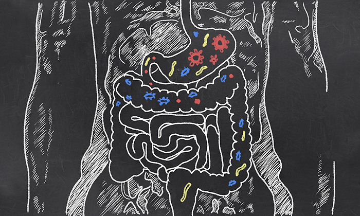
Far more than a simple tube for absorbing nutrients, the gut has three major constituents: luminal microbiota with an outer mucus layer, an inner mucus layer, and finally the epithelial layer. The luminal microbiome within the gut has around one thousand grams of bacteria that digest nutrients to produce hormones and vitamins, inhibit growth of pathogenic organisms, and assist in the metabolism of drugs and toxins.4-6 The intestinal cells known as enterocytes ultimately prevent substances from coming in contact with the blood and adaptive immune system through their tight intercellular junctions.4 When functioning normally, this barrier is highly selective, allowing nutrients to pass but still protecting against foreign molecules and pathogens.
Certain triggers and stresses are thought to cause dysbiosis and cellular junction disruption allowing for increased intestinal permeability, which has been implicated in the pathogenesis of many disease states including inflammatory bowel disease, celiac disease, hepatic fibrosis, food intolerance, and small intestinal bacterial overgrowth.7-9 Interestingly, there are also non-gastrointestinal diseases on this list, including fibromyalgia, chronic fatigue syndrome, and, relevant to dermatology, atopic dermatitis.10
The exciting developments in understanding of filaggrin and related proteins in atopic dermatitis (AD) have led to a deeper understanding of the central importance of barrier function in this disease.11 Along with skin barrier dysfunction, it has been postulated that there is compromise of the intestinal permeability barrier in atopic dermatitis as well. Many clinicians employ elimination diets—ostensibly in hopes of avoiding food allergens or triggers—and often report positive results from their afflicted patients despite fairly convincing evidence that dietary exclusion does not seem to bene t unselected patients.12 Directly or indirectly, the implication of the gut in AD has been very difficult to shake, with patients often insisting diet is the “root cause.”13
One of the ways that the intestinal barrier can be tested is to administer a mixture of sugars orally, frequently lactulose and rhamnose or mannitol, and examine their urinary excretion. These water-soluble, non-degradable molecules are instructive as the larger molecules (lactulose) are thought to permeate between the cells only when there is significant gut barrier impairment, while the smaller molecules (rhamnose or mannitol) are absorbed transcellularly, relatively independent of the barrier function. Therefore, by examining what is excreted in the urine, one can determine a lactulose/rhamnose ratio, which is elevated in proportion to gut barrier dysfunction.14
Compellingly, a study from the United Kingdom identified significantly increased lactulose to rhamnose excretion ratios (leaky gut) in children with atopic dermatitis when compared to controls, and were particularly greater in children under the age of eight years old.15 To examine the relationship between leaky gut, atopic dermatitis, and cow’s milk allergy, one study consisting of 35 infants under the age of one year with atopic dermatitis examined the urinary concentrations of end-products in children with cow’s milk allergy and AD compared to children with atopic dermatitis alone. Perhaps as one might expect, children with both cow’s milk allergy and atopic dermatitis had higher levels of the end-products, indicating greater intestinal permeability, compared to children with atopic dermatitis only.21
Another very recent study found a strong correlation between a gut bacteria called Faecalibacterium prausnitzii and atopic dermatitis, and that these patients had markers of gut epithelial inflammation, which can lead to barrier impairment.10 While these represent correlative data, they raise the important question of the clinical implication of leaky gut in these patients, particularly as we learn more about the role of transcutaneous sensitization in atopic dermatitis, secondary to what might be dubbed “leaky skin.”16
Armed with these fundamentals, we now move into the more clinically relevant realm: If leaky gut is indeed involved in the pathogenesis of atopic dermatitis, can it be treated? The answer seems to be “yes,” although with qualifications.
Diet has been implicated in the development of both leaky gut and atopic dermatitis and is a provocative place to start. Parents often note an association between certain foods and flares of atopic dermatitis.17 When tested, some 39 percent of families recognized improvement in their children with AD following an elimination diet, although this remains a confusing and controversial area.18 Historically, the thinking has been that food allergies (present in about 30 percent of children with atopic dermatitis versus only 10 percent in non-atopics) may be driving the skin disease.18
Indeed, food allergies have been shown to cause inflammation within the gut, altering the complex intestinal barrier. One small study evaluated 15 children with atopic dermatitis and subjected them to a two-week elimination diet where they were only allowed to consume a highly restricted group of foods. Interestingly, nine of the 15 patients had a significant reduction in clinical eczema scores and mean intestinal permeability in responders was lower when compared to that of non-responders.20 While studies suggest that only 40 percent of children with atopic dermatitis that are food sensitized actually have specific signs and symptoms of food allergy, these findings raise the idea that perhaps there are other mechanisms at play beyond simple IgE-mediated allergic responses.17
While many questions remain unanswered about diet and AD, there is hope that better understanding will help sort what types of dietary modifications could be helpful and in what settings.
Greater knowledge of the gut-skin connection has led to introduction of probiotic therapy in the management of atopic dermatitis. This concept relies on gut biodiversity and/or the presence of certain beneficial organisms as critical components to immunologic well being.
Perturbations in gut microbacterial diversity may inhibit the maturation of the T-regulatory cells causing Th1 and/ or Th2 induced in ammation.22-23 While the overall body of data on probiotics and atopic dermatitis remains somewhat unclear, the consensus currently states that probiotics are not effective for the treatment of established atopic dermatitis but may be helpful in prevention. However, there is the suggestion that the non-uniformity of the results from the many studies belies a more complex answer: not as simple as “probiotics are not effective”, but that there may be different phenotypes of the disease which respond more favorably than others; depending on the preponderance of the phenotype studied, the results could vary greatly.24
One double-blind placebo controlled crossover study administered two different strains of lactobacilli probiotic to 41 children with moderate to severe atopic dermatitis for six weeks. Ultimately, 14 patients had lactulose-mannitol excretion tests after placebo, and after active treatment to assess intestinal permeability. Median lactulose excretion decreased by 50 percent following treatment with probiotics, suggesting a significant improvement in gut barrier.24 Another study of 39 infants with atopic dermatitis were randomized to receive lactobacillus rhamnosus GG for three months, and the major groups of gut and skin bacteria were characterized using PCR along with several immune indices. At one month, the proportion of IgA- and IgM-secreting cells decreased significantly in the treatment group, thought to indicate an improved gut barrier function in those patients who received the probiotic.26
It is important to note that many unknowns remain here: the appropriate strain, dose, and duration of probiotics for atopic dermatitis still require a great deal of elucidation, but there is certainly promise in probiotic therapy.
Newer physical modalities may also be of benefit in conditions that result from possible compromise of the gut barrier. Gelatin tannate is one such agent that, through its chemical structure, may be able to form bonds with mucin within the mucus layers of the gut barrier.4 In addition to its physical activity in preventing contact of commensal organisms with the adaptive immune system, gelatin tannate acts as an astringent and can complex pro-inflammatory mucus-related proteins for elimination.25 This may prove to be a viable option for treating impaired barrier function, perhaps akin to a topical moisturizer for the skin.29
Atopic dermatitis remains a complex, multi-faceted disease. Despite its increasing prevalence and concomitant growing scientific interest, there are still many unanswered questions. Leaky gut represents a thought-provoking notion that has diagnostic and therapeutic rami cations, but raises many questions. More studies are required to determine if leaky gut is universal in atopic dermatitis or if there are specific subtypes/phenotypes for which it is relevant, and what approaches will yield the most improvement. Until then, as with all poorly-understood entities, we must keep alert for connections and observations that will guide future questions and research directions, and perhaps most of all, keep listening to our patients.
 Peter A. Lio, M.D. is Assistant Professor of Clinical Dermatology and Pediatrics-Dermatology at Northwestern University Feinberg School of Medicine and Director of the Chicago Integrative Eczema Center. Dr. Lio is also a member of the National Eczema Association Scientific Advisory Committee.
 Karan Lal, MS-4 is at New York Institute of Technology College of Osteopathic Medicine.
Sources
Original article written by Peter A. Lio and Karan Lal, MS-4 for Practical Dermatology. Republished with permission.
Learn what’s new and exciting in eczema research from the National Eczema Association research team.

Dermatologists weigh in on the benefits of LED face masks for eczema.

Learn why eczema often changes during puberty, common triggers for teens and how to minimize flares and find relief.

Jaylin Anderson, from Mason City, Iowa, shares her experience of trying to get her daughter’s eczema diagnosed and treated when she was an infant.


Evidence-based articles, expert-sourced lifestyle tips and stories from your community.