People of all skin colors, races and ethnicities can be affected by eczema, yet much of what is currently known has been learned by studying eczema in white skin.1 Recent studies of the high eczema prevalence in diverse racial and ethnic groups has resulted in calls for more studies on eczema on darker skin or skin of color (SOC).2 It is also crucial for diverse patient groups to participate in clinical trials for the rapidly growing treatment options to understand the various drugs’ real world applications for all eczema patients. Here we review what is currently known about eczema in SOC and highlight areas of focus for addressing gaps in research and enhancing the care of eczema patients with SOC.
What is meant by “skin of color”
The true number of skin colors is unknown and may be infinite.3 Historically, however, one way healthcare providers and scientists have referred to different skin tones is through use of a numeric scale called the Fitzpatrick scale (Figure 1).4 Developed in 1975, the scale was originally created to help physicians understand how much ultraviolet light combined with photo chemotherapy (PUVA) should be given to different patients during phototherapy to treat psoriasis.5 Dr. Fitzpatrick initially defined a I-IV scale based on patients with white skin reporting redness and tanning reactions to their first summer sun exposure. Type I was “always burn, never tan” and type IV was “rarely burn, tan with ease” with “usually” or “sometimes” burn in between. Types V and VI for brown skin and black skin were added later based on studies of pigmentation after ultraviolet light exposure.6,7
Using this scale, SOC is often referred to as types Types IV-VI, although individuals with SOC can identify with other Fitzpatrick skin types, highlighting the limitations of this scale.8,9 While the Fitzpatrick scale was not designed to be used to discuss race or ethnicity, a recent report suggests this does occur in some clinical settings.8,9 The most important take-home is that a “erythema” (redness) reaction to ultraviolet light does not equate to the degree of pigmentation in the skin. A more contemporary and racially and ethnically inclusive definition of SOC was created by the Skin of Color Society to include individuals of Asian, Hispanic/Latino, African, Native American, Pacific Island descent and people of mixed descent.
New methods to objectively quantify skin color using technology called “colorimetry” and “spectrophotometry” which measure the degree of pigmentation (white skin to brown skin) and hemoglobin (red) are being investigated to help give researchers and clinicians a unifying language with which to discuss SOC.10
Prevalence of eczema in skin of color
While eczema is a common skin disease, determining the prevalence of eczema in SOC in racial and ethnic populations is difficult, as published studies have not uniformly defined race and ethnicity categories. Terms used to define groups with regard to race/ethnicity have included White, Black, Hispanic, Hispanic origin, non-Hispanic, African American, Asian or Pacific Islander, American Indian, Puerto Rican, Mexican, Mexican American, Cuban or Cuban American, Dominican, Central or South American, multiracial, multiple races, stratifications by country of origin without reference to skin color and others.11-16 Despite this inconsistency across studies and gaps in data for certain SOC populations,17 the current understanding of eczema prevalence in SOC has pointed to several key observations:
At least 1 in 10 individuals are affected by eczema over a lifetime representing all races and ethnicities.14,16,18-20
Although study percentages vary, adults that are multiracial or white tend to have the highest prevalence of eczema.12,16,21,22
In the U.S., eczema affects more Black children (about 20%) than white children (about 16%) or Hispanic children (about 8%).23
Black children report more visits to healthcare providers and more prescriptions compared to white children,24 and Black race is associated with higher eczema-related out of pocket costs for eczema.25
Black children and Hispanic children tend to have more severe eczema than white children.22,26
Understanding differences in eczema diagnoses, symptoms, severity and treatment based on lighter skin versus darker skin or race or ethnicity definitions is challenging. Dr. Zelma Chiesa Fuxench, M.D., of the University of Pennsylvania said, “My impression is that we have a long way to go to fully understand the burden of atopic dermatitis (AD) across patients in the SOC spectrum. We need more studies that focus on the epidemiology of this disease among SOC patients to truly understand how it compares to better studied populations. Not only should these studies be examining the prevalence of AD but also the distribution of disease severity, and presence of comorbid atopic and non-atopic diseases. Another topic of interest would be the disparities in access to care, use of emergency and urgent care services, lack of access to dermatologist and treatment access among SOC patients with AD.” Dr. Andrew Alexis, MD, MPH, Chair of Dermatology and Director of the Skin of Color Center at Mount Sinai agreed, stating that “The majority of the world’s population can be characterized as having SOC and by the year 2044 more than half of the United States population will belong to a non-white racial and/or ethnic group. Clearly more studies are needed to understand eczema in SOC.”
Contributing factors to eczema: what is known in SOC
Skin barrier
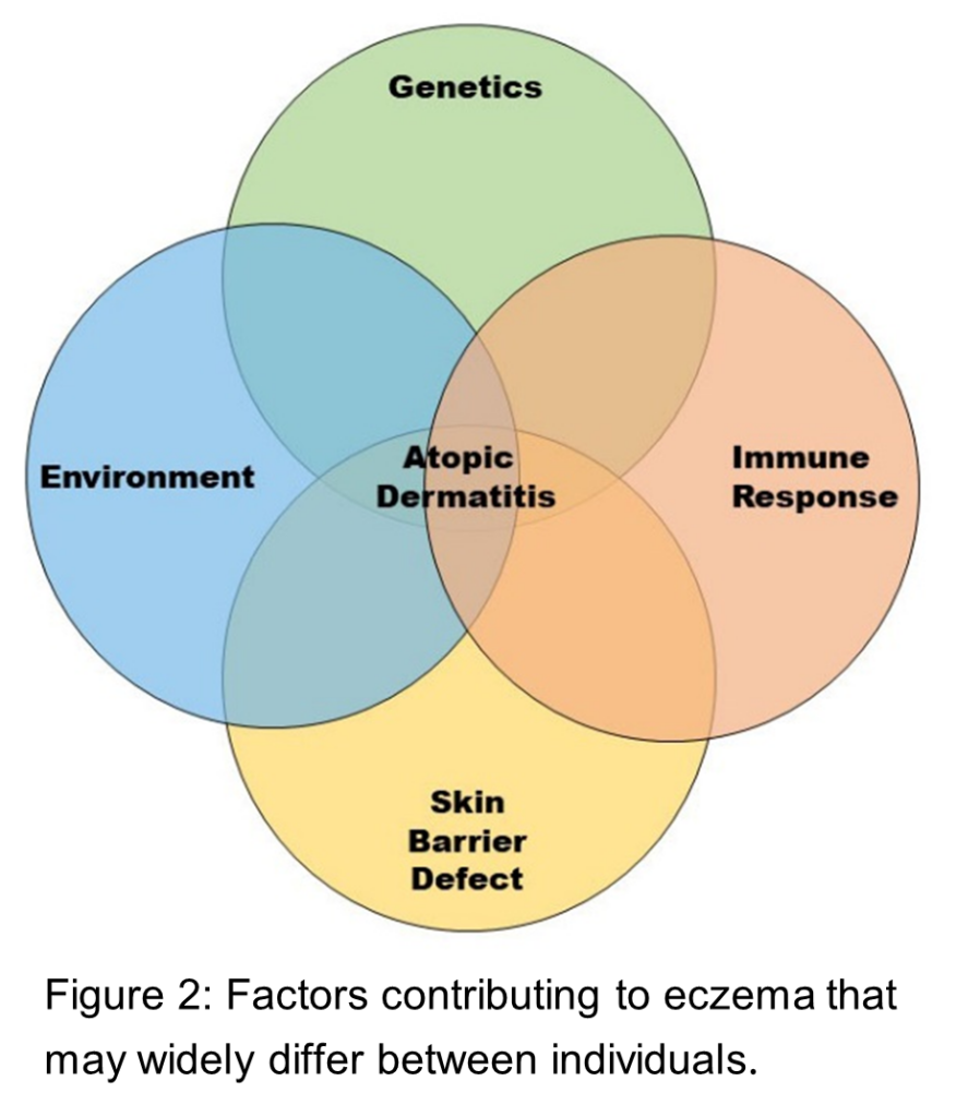
Eczema is caused by a combination of factors including a defective skin barrier, genetics, each person’s immune makeup and response and environmental exposures (Figure 2).
The impaired skin barrier in eczema results in higher levels of water loss from the skin (trans-epidermal water loss or TEWL). Some studies have found that TEWL is greater in Black skin compared with white skin. Further, a study including Asian, African and Danish individuals (total n=71) found significant differences in the fat (lipid) content of the outer layer of the skin that protects against water loss (the stratum corneum), with Asians having the highest lipid content, Danish having intermediate values and Africans having the lowest values.27 While studies are limited they suggest there may be racial variations in the skin barrier that contribute to eczema.
Genetic factors
The most common genetic mutation associated with susceptibility to eczema is filaggrin (FLG) whose protein product is responsible for binding specialized skin cells called keratinocytes together to create the structure of the stratum corneum. More than 300 different kinds of mutations within the FLG gene have been found and more than 20 of them have been associated with susceptibility to eczema.28 Four FLG mutations are consistently associated with eczema in patients of European ancestry and these have been the most commonly studied mutations. Studies of patients with East Asian ancestry show that there are more kinds of FLG mutations but only one has been associated with eczema in Chinese, Japanese, Korean and Taiwanese populations.28 Dr. Chiesa Fuxench says, “With respect to improving our understanding of the pathophysiology of AD, studies are needed that examine the genetics of AD across diverse populations. The FLG gene is the strongest known genetic risk factor for AD. However, most studies have primarily focused on those of European or Asian descent with few studies focused on those of African or Latinx ancestry.” Surprisingly, several studies did not detect FLG mutations in people with African ancestry until Dr. David Margolis, MD, PhD, of the University of Pennsylvania and colleagues studied 262 American children of African descent and found that 12.2% had FLG mutations of any type, nine different FLG variants were detected and those nine variants associated with more persistent eczema than children with the normal FLG gene.28 A subsequent study showed that FLG variants in African Americans correlated with African FLG variants and FLG variants in European Americans correlated with European FLG variants despite the mixture of the two populations in America.29 Overall, the number of FLG variants differs between African Americans and European Americans,29 yet a recent study found these differences do not appear to explain AD risk between these racial groups.30
Immune system factors
The last several years have seen an expansion of studies investigating the complex immune pathways underlying AD, and more specifically what immune pathways are regulating AD in different populations. An initial study looked at the types of T helper (Th) cells, a type of immune cells, in the skin of Asian (Japanese and Korean) AD populations compared to European American AD patients.31 Japanese and Korean AD patients had higher numbers of certain types of T helper cells (Th17 and Th22) that cause inflammation than European American AD patients.31 A further study then looked at the T cell-produced messengers (cytokines and interleukins) in the blood of Japanese and Korean vs. European American patients, finding important differences in these messengers between groups.32 A third study comparing Han Chinese with European American AD patients again found higher numbers of Th17 inflammation-causing cells in Chinese patients.33 Comparing African American with European American AD patients, researchers found lower numbers of certain Th1/Th17 inflammation-causing cells and higher numbers of other inflammation-causing cells (Th2/Th22) in the affected skin from African American patients.33 As new therapies for AD targeting different immune pathways are developed, understanding immune system similarities and differences between racial or ethnic people groups will help guide a more targeted treatment selection approach for diverse eczema patients.
Treatment of eczema in SOC
The first step toward treatment of eczema in SOC patients is appropriate diagnosis, yet this can be challenging. Dr. Alexis said, “There are major differences in the appearance of eczema in SOC. The severity of eczema in SOC can be easily underappreciated since we do not really see the bright red color associated with eczema in white skin, the colors can range from gray to reddish brown to purple or purplish gray. Beyond color, another example is called the follicular pattern – tiny bumps which are extremely itchy and might correspond with hair follicles. Eczema in SOC may also appear to other types of skin diseases, leading to misdiagnosis.”
Based on the physical characteristics of SOC that may impact the barrier as discussed above, SOC is often more dry than white skin.34 Efforts are being made to analyze what types of moisturizers work best to treat dry skin in different populations including those with SOC, to enhance this cornerstone of AD treatment.34
Individuals with SOC can effectively use all available therapies for AD, however in some cases additional considerations may be needed. For example, as SOC contains more skin pigment that acts as a UV filter, higher phototherapy doses may be needed and there may be longer times before treatment efficacy. An objective system has not yet been established to predict UV light responses in SOC since the Fitzpatrick scale was not developed for SOC, so determining optimal dosing for these patients can be difficult.3 Patients with SOC can experience post-inflammatory pigmentation changes, both hyperpigmentation (skin darkening) and hypopigmentation (skin lightening).35,36 More studies are needed to address how pigmentation is impacted with treatment of AD in SOC.
Finally, the appearance of AD lesions and erythema (redness) in SOC can impact the assessment of treatment outcomes. Often “redness” is evaluated as part of the scoring system for clinical trials and to evaluate how well a drug is working for someone’s eczema, but “redness” can appear as pink, brown or purple depending on different skin color tones. A study from 2015 tested four visual AD outcome measures assessments (Eczema Area and Severity Index (EASI), objective-SCORing Atopic Dermatitis (oSCORAD), Three Items Severity index (TIS) and Six Areas, Six Sites Atopic Dermatitis (SASSAD) in 18 patients with various levels of skin darkness (African, Asian, Caucasian and Indian descent). The outcome measures were not as reliable and had reduced validity in highly pigmented patients because of the varying ability of healthcare providers to judge redness in certain skin colors.37 A subsequent study including 11 light-skinned patients and 14 patients with SOC evaluated EASI, oSCORAD, Investigator’s Global Assessment (IGA) and a novel greyscale that assessed clinical-sign greyness, graded from 0 to 3 (no grey = 0 to dark grey = 3) based on the average greyness of the affected area rather than redness. In this study EASI was found to be reliable in all skin colors and was proposed as the best outcome measure for AD clinical trials globally.38 In one study, after adjusting for the “redness score” in the oSCORAD scale, Black children were found to have six times greater risk of severe AD compared to white children, indicating that use of this redness score can lead to underestimating the severity and treatment outcomes of AD in Black children.39,40 Improved training on how skin diseases appear in SOC and greater inclusion of people with SOC in the validation of outcome measures for clinical trials will help improve the utility of these assessments in the future, influencing both diagnosis of eczema symptoms and treatment of eczema in SOC.
Efforts to address SOC underrepresentation and disparities in eczema knowledge and treatment
An important acknowledgement across medicine is that there has been a significant gap in representation of SOC in medical literature, educational materials, clinical trials and healthcare providers.4,41-43 Researchers recently analyzed the diversity in medical photographs from six plastic surgery journals and the New England Journal of Medicine over the past 30 years. Among the 24,209 color photographs depicting skin, 78% were of white skin and only 22% were of non-white skin.4 Of four main textbooks traditionally used to train U.S. dermatologists to diagnose skin diseases, the majority of images were of white skin, and in all cases the percentage of images depicting darker skin were not representative of U.S. populations.43 A systematic review of 58 U.S. dermatological clinical trials that recorded race/ethnicity found that around 75% of participants were white.44 Lastly, the representation of SOC among dermatological healthcare providers is behind the increasing U.S. population trends with 3% Black and 4.2% Hispanic dermatologists in the workforce compared to 12.8% Black and 16.3% Hispanic individuals in the general U.S. population.42
Importantly, there are individuals and organizations now working to increase knowledge about SOC and improve representation of images in the medical literature, including skin diseases like eczema.45 New guidance for proper lighting for medical images of SOC was recently published in the British Journal of Dermatology with the goal of increasing the number of quality of SOC images in educational and medical literature.46 Dr. Nada Elbuluk, MD, at the Keck Hospital of the University of Southern California said, “New textbooks and educational resources are being developed and the American Association of Dermatology is working on a SOC curriculum. Journals are diversifying their editorial boards and the major meetings of the scientific dermatology societies are consciously selecting more diverse content of what is presented.”
There is also great need for dermatologists with expertise in SOC. To address health disparities and issues of access to healthcare, SOC clinics are opening around the country. The first of these was the SOC Center at Mount Sinai St. Luke’s in New York City founded by Dr. Susan Taylor, MD, growing to 15 different centers by 2020.47 The goals of these centers are to: 1) further the care and research of conditions common among persons of color; 2) prepare residents to treat skin of color; and 3) educate the medical community and public.47 The Skin of Color Society, established in 2004, additionally drives this movement by promoting research and education of fellows, associate members, residents, and research fellows through grants, mentorship and industry partnerships. Dr. Elbuluk also directs the Skin of Color and Pigmentary Disorders Program to promote research and education and said, “The field of dermatology is underutilized as a specialty by people of color. They feel that they are being misdiagnosed and undertreated. Skin diseases in skin of color have to be approached differently. At these SOC clinics, patients of color can come and feel like people there have expertise and cultural understanding. I wanted a place that would change the narrative, where patients really felt heard and could see that their conditions were getting better.” With these dedicated clinics, and broader efforts to address knowledge and research gaps related to SOC, the diagnosis, care and treatment of eczema in SOC is poised to dramatically improve. Efforts to address and reduce the gaps should parallel the evolving racial/ethnic make-up of the AD community and the U.S. population.
Take home points:
Much of what is currently known about eczema has been learned by studying white skin. Healthcare providers and researchers are acknowledging and addressing these gaps to improve knowledge and care of eczema in SOC.
Skin, genetic and immunologic factors that contribute to eczema can differ between racial and ethnic groups and impact prevalence, persistence, and severity of eczema in SOC.
Increasing representation of SOC in medical education, medical literature, and the healthcare workforce, including specialized clinical centers, will improve healthcare for people with SOC.
References:
1. Roh YS, Huang AH, Sutaria N, et al. Real-world comorbidities of atopic dermatitis in the U.S. adult ambulatory population. J Am Acad Dermatol. 2021.
2. Montgomery SN, Elbuluk N. A quantitative analysis of research publications focused on the top chief complaints in skin of color patients. J Am Acad Dermatol. 2020.
3. Ware OR, Guiyab J, Okoye GA. Phototherapy in Skin of Color. Dermatol Clin. 2020;38(1):63-69.
4. Massie JP, Cho DY, Kneib CJ, et al. Patient Representation in Medical Literature: Are We Appropriately Depicting Diversity? Plast Reconstr Surg Glob Open. 2019;7(12):e2563.
5. Fitzpatrick T. Soleil et peau. J Med Esthet. 1975;2:33034.
7. Pathak M, Jimbow K, Szabo G, Fitzpatrick TB. Sunlight and Melanin Pigmentation. In: Smith K, ed. Photochemical and Photobiological Reviews. Vol 1. New York: Plenum Press; 1976:211-239.
8. Pichon LC, Landrine H, Corral I, Hao Y, Mayer JA, Hoerster KD. Measuring skin cancer risk in African Americans: is the Fitzpatrick Skin Type Classification Scale culturally sensitive? Ethn Dis. 2010;20(2):174-179.
9. Ware OR, Dawson JE, Shinohara MM, Taylor SC. Racial limitations of fitzpatrick skin type. Cutis. 2020;105(2):77-80.
10. Ly BCK, Dyer EB, Feig JL, Chien AL, Del Bino S. Research Techniques Made Simple: Cutaneous Colorimetry: A Reliable Technique for Objective Skin Color Measurement. J Invest Dermatol. 2020;140(1):3-12 e11.
11. Barbarot S, Auziere S, Gadkari A, et al. Epidemiology of atopic dermatitis in adults: Results from an international survey. Allergy. 2018;73(6):1284-1293.
12. Chiesa Fuxench ZC, Block JK, Boguniewicz M, et al. Atopic Dermatitis in America Study: A Cross-Sectional Study Examining the Prevalence and Disease Burden of Atopic Dermatitis in the U.S. Adult Population. J Invest Dermatol. 2019;139(3):583-590.
13. Hanifin JM, Reed ML, Eczema P, Impact Working G. A population-based survey of eczema prevalence in the United States. Dermatitis. 2007;18(2):82-91.
14. Shaw TE, Currie GP, Koudelka CW, Simpson EL. Eczema prevalence in the United States: data from the 2003 National Survey of Children’s Health. J Invest Dermatol. 2011;131(1):67-73.
15. Silverberg JI, Barbarot S, Gadkari A, et al. Atopic dermatitis in the pediatric population: A cross-sectional, international epidemiologic study. Ann Allergy Asthma Immunol. 2021;126(4):417-428 e412.
16. Silverberg JI, Hanifin JM. Adult eczema prevalence and associations with asthma and other health and demographic factors: a U.S. population-based study. J Allergy Clin Immunol. 2013;132(5):1132-1138.
17. Price KN, Krase JM, Loh TY, Hsiao JL, Shi VY. Racial and ethnic disparities in global atopic dermatitis clinical trials. Br J Dermatol. 2020;183(2):378-380.
18. Abuabara K, Magyari A, McCulloch CE, Linos E, Margolis DJ, Langan SM. Prevalence of Atopic Eczema Among Patients Seen in Primary Care: Data From The Health Improvement Network. Ann Intern Med. 2019;170(5):354-356.
19. Al-Naqeeb J, Danner S, Fagnan LJ, et al. The Burden of Childhood Atopic Dermatitis in the Primary Care Setting: A Report from the Meta-LARC Consortium. J Am Board Fam Med. 2019;32(2):191-200.
20. Silverberg JI. Public Health Burden and Epidemiology of Atopic Dermatitis. Dermatol Clin. 2017;35(3):283-289.
22. Chung J, Simpson EL. The socioeconomics of atopic dermatitis. Ann Allergy Asthma Immunol. 2019;122(4):360-366.
23. Fu T, Keiser E, Linos E, et al. Eczema and sensitization to common allergens in the United States: a multiethnic, population-based study. Pediatr Dermatol. 2014;31(1):21-26.
24. Fischer AH, Shin DB, Margolis DJ, Takeshita J. Racial and ethnic differences in health care utilization for childhood eczema: An analysis of the 2001-2013 Medical Expenditure Panel Surveys. J Am Acad Dermatol. 2017;77(6):1060-1067.
25. Chovatiya R, Begolka WS, Thibau IJ, Silverberg JI. Financial burden and impact of atopic dermatitis out-of-pocket healthcare expenses among black individuals in the United States. Arch Dermatol Res. 2021.
26. Silverberg JI, Simpson EL. Associations of childhood eczema severity: a U.S. population-based study. Dermatitis. 2014;25(3):107-114.
27. Jungersted JM, Hogh JK, Hellgren LI, Jemec GB, Agner T. Ethnicity and stratum corneum ceramides. Br J Dermatol. 2010;163(6):1169-1173.
28. Margolis DJ, Mitra N, Gochnauer H, et al. Uncommon Filaggrin Variants Are Associated with Persistent Atopic Dermatitis in African Americans. J Invest Dermatol. 2018;138(7):1501-1506.
29. Zhu Y, Mitra N, Feng Y, Tishkoff S, Hoffstad O, Margolis D. FLG Variation Differs between European Americans and African Americans. J Invest Dermatol. 2021.
30. Fulton RL, Margolis DJ, Sockler PG, Mitra N, Wong X, Common JE. No Association of filaggrin copy number variation and atopic dermatitis risk in White and Black Americans. Exp Dermatol. 2021.
31. Noda S, Suarez-Farinas M, Ungar B, et al. The Asian atopic dermatitis phenotype combines features of atopic dermatitis and psoriasis with increased TH17 polarization. J Allergy Clin Immunol. 2015;136(5):1254-1264.
32. Wen HC, Czarnowicki T, Noda S, et al. Serum from Asian patients with atopic dermatitis is characterized by TH2/TH22 activation, which is highly correlated with nonlesional skin measures. J Allergy Clin Immunol. 2018;142(1):324-328 e311.
33. Czarnowicki T, He H, Krueger JG, Guttman-Yassky E. Atopic dermatitis endotypes and implications for targeted therapeutics. J Allergy Clin Immunol. 2019;143(1):1-11.
34. Wan DC, Wong VW, Longaker MT, Yang GP, Wei FC. Moisturizing different racial skin types. J Clin Aesthet Dermatol. 2014;7(6):25-32.
35. Mitchell KN, Tay YK, Heath CR, Silverberg NB. Review article: Emerging issues in pediatric skin of color, Part 2. Pediatr Dermatol. 2021;38 Suppl 2:30-36.
36. Woolery-Lloyd H, Kammer JN. Treatment of hyperpigmentation. Semin Cutan Med Surg. 2011;30(3):171-175.
37. Zhao CY, Wijayanti A, Doria MC, et al. The reliability and validity of outcome measures for atopic dermatitis in patients with pigmented skin: A grey area. Int J Womens Dermatol. 2015;1(3):150-154.
38. Zhao CY, Hao EY, Oh DD, et al. A comparison study of clinician-rated atopic dermatitis outcome measures for intermediate- to dark-skinned patients. Br J Dermatol. 2017;176(4):985-992.
39. Ben-Gashir MA, Hay RJ. Reliance on erythema scores may mask severe atopic dermatitis in black children compared with their white counterparts. Br J Dermatol. 2002;147(5):920-925.
40. McKenzie S, Brown-Korsah JB, Syder NC, Omar D, Taylor SC, Elbuluk N. Variations in Genetics, Biology, and Phenotype of Cutaneous Disorders in Skin of Color. Part II: Differences in Clinical Presentation and Disparities in Cutaneous Disorders in Skin of Color. J Am Acad Dermatol. 2022.
41. Ly DP, Jena AB. Trends in Diversity and Representativeness of Health Care Workers in the United States, 2000 to 2019. JAMA Netw Open. 2021;4(7):e2117086.
42. Pandya AG, Alexis AF, Berger TG, Wintroub BU. Increasing racial and ethnic diversity in dermatology: A call to action. J Am Acad Dermatol. 2016;74(3):584-587.
43. Reilley-Luther J, Cline A, Zimmerly A, Azinge S, Moy J. Representation of Fitzpatrick skin type in dermatology textbooks compared with national percentiles. Dermatol Online J. 2020;26(12).
44. Charrow A, Xia FD, Joyce C, Mostaghimi A. Diversity in Dermatology Clinical Trials: A Systematic Review. JAMA Dermatol. 2017;153(2):193-198.
45. Aoki V, Oliveira M, Wegzyn C, et al. Assessment and Monitoring Challenges Among Patients With Moderate-to-Severe Atopic Dermatitis Across Fitzpatrick Skin Types: A Photographic Review and Case Series. Dermatitis. 2022.
46. Lester JC, Clark L, Jr., Linos E, Daneshjou R. Clinical photography in skin of colour: tips and best practices. Br J Dermatol. 2021.
47. Tull RZ, Kerby E, Subash JJ, McMichael AJ. Ethnic skin centers in the United States: Where are we in 2020? J Am Acad Dermatol. 2020;83(6):1757-1759.
Eczema isn’t just a condition you manage, it’s something you live with every day. It shows up in quiet moments and public ones, in routines, relationships and everyday decisions. It can mean planning around flare-ups…
Today the eczema community is one step closer to having a new treatment option. The U.S. Food and Drug Administration (FDA) has approved ANZUPGO® (delgocitinib) cream, from manufacturer LEO Pharma Inc., as the first topical…
Learn what’s new and exciting in eczema research from the National Eczema Association research team.
Get the latest eczema news delivered to your inbox.
Advances in research and treatments
Lifestyle tips and hacks
Stories from the community
NEA Newsletter Signup with Segmentation
Please support this website by adding us to your whitelist in your ad blocker. Ads are what helps us bring you premium content! Thank you!
We use technologies such as cookies in order to integrate with social media, serve relevant advertising, analyze our traffic and improve and customize your experience on our websites. By continuing to use this site, you consent to our use of cookies. Privacy Policy
The latest in eczema news, delivered
Evidence-based articles, expert-sourced lifestyle tips and stories from your community.